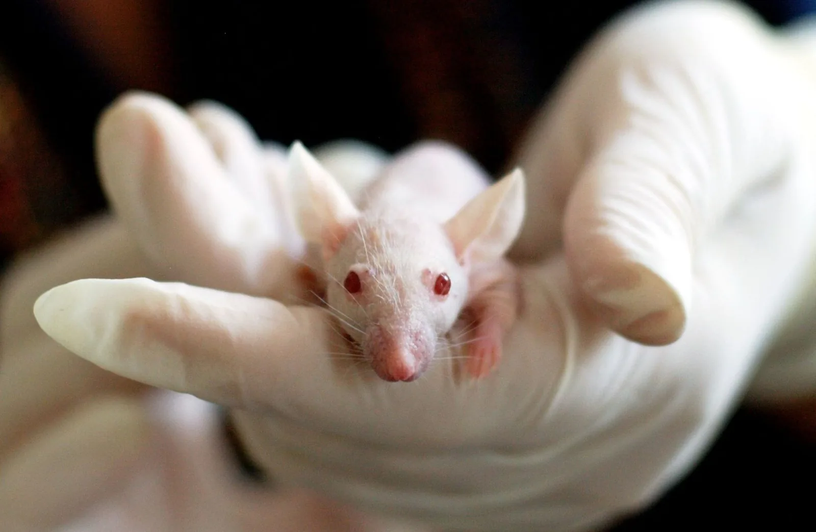
In September 2026, a photograph from the Pașca lab at Stanford University began making the rounds. It showed a cross-section of a mouse brain with dashed white lines drawn around a cavity that should not have been there. The label said "xenocortical graft." Within a day, "xenocortical" had jumped from a caption in Nature to the front page of The Guardian. The word is new. The idea it describes is one of the most striking in modern biology: a mouse whose brain contains living, functioning human cortical tissue.
Breaking the word apart
"Xenocortical" is built from two pieces. Xeno- comes from the Greek for "foreign," the same root as xenotransplantation — moving tissue from one species into another. Cortical refers to the cortex, the deeply folded outer sheet of the brain where perception, language, planning, and self-awareness are thought to arise. A xenocortical mouse is, precisely: a mouse that has received a transplant of cortical tissue from another species — in this case, human tissue grown from stem cells in a dish.
How the animals are made
The recipe, as described in the Nature paper and its coverage, runs like this:
- Start with a patient's skin cells. These are reprogrammed back into stem cells, then guided to become neural tissue — so-called brain organoids, each containing roughly 100,000 cells at the time of injection.
- Make room. The recipient mice are genetically engineered so that their own cortex and hippocampus barely develop, leaving a cavity in the brain.
- Inject the human organoids into that cavity.
- Wait. Three months later, the graft has hooked itself into the mouse's blood supply, filled the empty space, and grown to around four million cells — making up roughly half the brain by volume.
What the human tissue actually does
The transplanted cells are not passengers. They connect to the mouse's own neurons, extend wiring toward the spinal cord, and respond to the animal's physiology. In one experiment, five hours of low oxygen selectively damaged the human nerve cells — a vulnerability that mirrors what happens in human cerebral palsy. The graft even contained von Economo neurons, the large spindle-shaped cells that are among the first to die in frontotemporal dementia. The human tissue behaves like human tissue.
What a xenocortical mouse is not
Just as important is what the paper insists these animals are not. The human tissue is not a miniature human brain; it is immature, described as roughly halfway through fetal development. It is not wired like a human cortex. The mice are not smarter — the researchers report only modest improvements in gait and cognition, and are careful to say the animals are not "enhanced." A xenocortical mouse is a model system: a living, vascularized, interactive version of the human brain tissue that scientists previously could only grow in a dish.
Sergiu Pașca's point: the human brain is inaccessible. You cannot biopsy a living person's cortex to study schizophrenia. A xenocortical mouse makes human brain development and function accessible for investigation.
Why the word matters
Naming a thing changes how people argue about it. "Humanized mouse" suggested a mouse with a few extra cells. "Xenocortical" names the specific, remarkable fact: foreign cortical tissue, integrated and alive, in another animal's head. It is the right word for a technology that has crossed a line — from growing human brain tissue in a dish to giving it a body — and it forces us to talk honestly about what comes next.
Sources
- Pașca, S. P., et al. (2026). Xenocortical transplantation of human cortical organoids in mice. Nature. doi:10.1038/s41586-026-11032-2
- Sample, I. (2026, September 16). "Scientists create mice with part-human brains." The Guardian.
- Revah, O., et al. (2022). Maturation and circuit integration of transplanted human cortical organoids. Nature, 611, 518–526.
- Mansour, A. A., et al. (2018). An in vivo model of functional and vascularized human brain organoids. Nature Biotechnology, 36, 432–441.